Sunday, May 5, 2013

Anti TB Treatment in India Needs to be Improved

Source: Anti TB Treatment in India Needs to be Improved


India has the highest burden of tuberculosis in world accounting for 20% of global incidence of TB. Recently, a survey of tuberculosis prescription practices in private and public sector in India was published in the current issue of National journal of integrated research in medicine by Dr. Gyanshankar Mishra from Nagpur, India. The study found that more than 90% prescriptions in the private sector and RNTCP (Revised National Tuberculosis Control Programme) were incorrect as per the WHO (World Health Organization) norms. The study further showed that factors for development of drug resistant TB were more in prescriptions by private practitioners whereas overdoses of anti TB medicines were more in RNTCP. The study notes that the patients above 30 kg in RNTCP receive the same dose of all anti TB drugs except Rifampicin. As a result a person weighing 30 kg receives 300 mg of INH whereas another person weighing 31 kg receives 600 mg of INH , thus receiving almost double the recommended dose of 10 mg per kg. The study has recommended creation of weight bands under RNTCP for persons weighing more than 30 kg. The study concludes that the anti TB treatment offered in private and public sector in India at present is not satisfactory and needs to be improved.
Link to article:

The abstract of this article is given as below.
Introduction: India has the highest burden of tuberculosis in world, accounting for 20% of global incidence of TB (Tuberculosis). TB treatment is available both in private and public sector in India.
Aim & objectives: The current study was carried out to study and compare the prescribing practices of anti-tuberculosis medications by private practitioners and healthcare providers in public sector (under RNTCP-Revised National Tuberculosis Control Programme).
Material and Methods: 105 anti TB prescriptions of private practitioners and 105 RNTCP (Revised National Tuberculosis Programme) treatment cards were analysed.
Results: 9.52% prescriptions by private practitioners and 4.76 % RNTCP prescriptions were correct. Factors for drug resistance were present in 67.62 % of prescriptions by private practitioners and 28.57 % of RNTCP prescriptions whereas overdosing was present in 53.33 % of prescriptions by private practitioners and 68.57 % of RNTCP prescriptions.
Conclusion: The anti TB treatment offered in private and public sector in India is not satisfactory at present and needs to be improved
Reference: Mishra G, Mulani J. Tuberculosis Prescription Practices In Private And Public Sector In India. NJIRM. 2013; 4(2): 71-78.

Saturday, April 13, 2013

Diabetes Interferes in Lung Functions of Asthma and COPD Patients

Diabetes Interferes in Lung Functions of Asthma and COPD Patients

Monday, March 12, 2012

Has anyone ever explained why you have COPD?

Has anyone ever explained why you have COPD?

Classic COPD appearences as pulmonary enphisema & Chronic bronchitis
Make the following experiment: Inhale as much as you can, fill your lungs with air and hold your breath. Now, exhale a little, and soon after hold your breath. From this point (even with lungs full of air), to breathe again, but based on this interval.
It is exactly this way that a COPD patient breathes with lungs full of air, but with an ability to Exhale (put the air out) Limited.
This is because COPD, an acronym for Chronic Obstructive Pulmonary Disease (more commonly known as emphysema or chronic bronchitis), causes an inflammation in the bronchi of the lungs that obstruct the air passage. As the air out of the lung must be “pushed” by a positive pressure generated by the muscles, he finds difficulty and became trapped, leads to hyperinflation, one of the reasons for the lack of air or oxygen.
COPD
COPD is a disease still largely unknown by most people, including their holders.
The profile of the person with COPD is generally a person smoking, about 40 years and that has as its main symptoms are cough and shortness of breath.
Smokers think the cough is a natural product that comes “free” with the cigarette and that one day will pass … Big mistake! The cough does not cease, by contrast, tends to worsen and the smoker becomes accustomed to thinking that the cough is normal!
Diagnosis of COPD
The vast majority of people who smoke because they think they made a chest x-ray recently, and this is with the report “normal.” Unfortunately the picture is not all! The chest radiograph may be normal, even though the patient may have COPD.
The lab exam that diagnoses COPD is spirometry or pulmonary function test (also known as “blow test”). The spirometry assesses lung function data that may indicate to be a smoker with COPD.
But remember: Smoking causes 52 different diseases and COPD is one of them!
Tobacco Industry & COPD smoker
For the “happiness” of tobacco industry, as cigarettes industries, smokers think they can quit at any time. This, for the vast majority of people do not happen, for one simple reason:

Cigarettes contain nicotine that is an addiction substance, provokes quimic dependency and makes very difficult to quit smoking!
Accordind to PULMAOSA Medical Editor, Dr. Marcos Nascimento: “Quit smoking without medical supervision is extremely difficult, only 3-5 people among 100 do. Hence the need to search for medical and psychological help to start a cognitive behavioral treatment, complement Dr. Nascimento. And if you need to use medication to alleviate withdrawal symptoms – which is that irrepressible desire to return to smoking, there are some medicines available today. Thus, the treatment of smokers have a greater chance of success and you can prevent the progression of COPD.
Tobacco Industries and its Marketing Policy
Like any commercial enterprise, tobacco industries target PROFIT, and just want to be able to pay their shareholders. Tobacco Industries obviously know that the customer can get sick! In fact, it’s just knowing that your customers will get sick and may die or cease to be customers, they have developed a highly focused marketing on young people and women to feed back their customer base and therefore continue their lucrative business.
There is a Chinese saying that someone asks when is the best time to plant a tree. And the answer says that the best time was twenty years ago. The second best time is now!
Therefore, so it is with COPD and smoking: The Best, would be no smoking at any time, but will ALWAYS be an excellent time to STOP SMOKING. Therefore, seek the help of a pulmonologist to informs you about the dangers of smoking and lead the best treatment for COPD.
Sources:
PULMAOSA- Lungs: Your Atmosphere, Your Life!

Wednesday, October 26, 2011

This Diwali remember – What you burn is what you breathe!


As sure as Diwali is a festival of lights, sweets, gaiety, splendor & fireworks, it is also one of deafening noise, blinding light, risky fire & suspended particles. And this has direct effects on our health & environment. When the entire nation looks forward to Diwali as a celebration of life, patients suffering from Asthma & COPD (Chronic Obstructive Pulmonary Disease) begin readying there lifesavers – Inhalers, nebulizers or whatever gives them a breath of life. For these people Diwali is not a festival of light & gaiety but that of smoke, coughing & wheezing.
Fireworks are sources of some of the highly toxic inhalants produced during Diwali celebrations. Firecrackers are “power packed” with potassium nitrate, carbon & sulfur. Apart from this they also contain toxic contents like copper, cadmium, lead, manganese, magnesium, zinc, sodium, potassium, and aluminum powder & barium nitrate.  When ignited, the crackers burst allowing these powerful chemicals to come in contact with atmosphere and the smoke thus generated contains increased amounts of carbon monoxide, carbon dioxide, nitrogen dioxide, hydrocarbons, hydrogen sulfide & particulate matter which worsen the quality of Diwali air. The suspended particles hog like a thick blanket reducing visibility and suffocating the atmosphere. In fact studies have demonstrated that during Diwali festival the concentration of sulfur dioxide increased by 10 times & that of nitrogen dioxide, PM10(Particulate matter of size less than 10 microns) & TSP(Total Suspended Particles) increased by 2 to 3 times. The Overall air pollution during Diwali increases by about 200 %. The biggest culprits among firecrackers are the colour sparkles (“Phuljari”), “Anar”, “Chakri”, Fire pencils, Snake tablets & “Hydrogen” bomb.
A special mention may be made of PM10. With the average PM10 charge, we inhale millions of fine particles with each breath. The larger particles( 5 to 10 microns) are filtered in the nose & throat, smaller particles (3 to 5 microns) arrive in the bronchial tube, bronchi (2 to 3 microns), bronchioles (1 to 2 microns) & in alveoli (0.1 to 2 microns) & finally in the blood. These particles can no longer be coughed up & as deposits lead in long term to inflammation, particularly in asthmatics, & also with healthy people, although they may not notice the immediate irritant effects.
When we inhale such a highly toxic and polluted air during Diwali, how can escape from its ill effects? The harmful oxides present in the Diwali fumes come into contact with the moisture while passage from the nostrils to the lungs & form acids which cause immense damage to the body.
The Diwali smoke potentially leads to development of various respiratory ailments like –Allergic bronchitis, acute exacerbation of bronchial asthma & COPD (Chronic Obstructive Pulmonary Disease), allergic rhinitis, laryngitis, sinusitis, pharyngitis, common cold, acute eosinophilic pneumonia, reactive airway dysfunction syndrome, etc.
Children, pregnant women, asthmatics & senior citizens are highly prone to these potentially harmful effects of Diwali smoke.
Here is a list of precautions that need to be followed during Diwali celebrations.
Precautions to be followed by asthmatics:
1.      Stay away from people burning crackers.
2.      Keep the inhalers and other medicines ready beforehand (both maintenance & reliever medications).
3.      Consult your doctor and start maintenance dose of inhalers a few days before the festival & continue the same two days after Diwali.
4.      In severe cases rescue medications need to be taken & if not relieved contact your doctor immediately.
5.      Better do not venture out in the evening of Diwali. Stay in company of friends and family members in house.
6.      If need to venture outside use masks (N95 masks have been technically recommended).
7.      Consult your doctor regarding pulmonary vaccination in advance.
8.      Stay away during colouring and white washing of house before Diwali because these also act as “triggers” of an asthma attack.
9.      If possible plan a visit to some hill-station / ecoclean place (which is not much crowded) during Diwali festival.
Precautions to be followed by every one of us during Diwali festival:
1.      Avoid /decrease the firework celebrations & play “ecosafe” Diwali.
2.      Enjoy Diwali with lamp, lanterns & diyas.
3.      Fireworks if carried out should be done in open grounds, away from residential areas & during fixed time limits.
We should remember that one person’s idea of fun could be an asthma patient’s nightmare. Asthma patients have as much right as us to stick around and enjoy Diwali. And ultimately for our own benefit we should not forget that “what we burn is what we breathe”, this Diwali.

Author:
Dr. Gyanshankar Mishra
MBBS, MD(Pulmonary Medicine), DNB(Respiratory Diseases)
Chest Physician, Nagpur
email address: chestcareclinic@gmail.com .

Sunday, August 29, 2010

Swine flu revised guidelines - India


So far, the present guidelines stipulate that a person suspected of influenza A H1N1 need to be referred to an identified govt. health facility. He/she needs to be kept in an isolation facility in that hospital and if found positive, is treated accordingly. 
In order to make the testing facility for H1N1 more accessible at large and due to the onset of the Influenza season in the country, it has been decided to revise the existing guidelines.
Under the new guidelines, any person with flu like symptoms such as fever, cough, sore throat, cold, running nose etc. should go to a designated Government facility for giving his/her sample for testing for the H1N1 virus.  After clinical assessment, the designated medical officer would decide on the need for testing. Except for cases that are severe, the patient would be allowed to go home (This was not allowed under the existing guidelines).    
The sample of the suspect case would be collected and sent to the notified laboratory for testing. If tested as positive for H1N1 and in case the symptoms are mild, the patient would be informed and given the option of admission into the hospital or isolation and treatment at his own home.
In case the patient opts for home isolation and treatment, he/she would be provided with detailed guidelines / safety measures to be strictly adhered to by the entire household of the patient.  He/ she would have to provide full contact details of his entire household. The house hold and social contacts would be provided with the preventive treatment.
Notwithstanding the above guidelines, the decision of the doctor of the notified hospital about admitting the patient would be final.

In case the test is negative, the patient will accordingly be informed.
These guidelines have been issued by the Government in public interest and shall be reviewed from time to time depending on the spread of the pandemic and its severity in the country.  These guidelines would however not apply to passengers who are identified through screening at the points of entry.  The existing policy of isolating passengers with flu like symptoms would continue.

Thursday, August 19, 2010

TB: Why you should not discriminate


Impact of Stigma and Discrimination

Tuberculosis is an infectious disease caused by bacteria that any person can get. It is not a hereditary disease or a curse of God. It can be completely cured by taking regular and complete treatment. Stigma and discrimination against people diseased with TB can occur in many settings at the workplace, health care facilities, or within the community. Its manifestation can be as dramatic as physical violence or as subtle as avoidance. However, it is totally unnecessary and primarily based on myths. Stigma is as old as history. Stigma and discrimination against people infected with TB can occur in many settings at the workplace, healthcare facilities, or within the community. Its manifestation can be as dramatic as physical violence or as subtle as avoidance. However, it is totally unnecessary and primarily based on myths. Beyond the economic consequences, stigma and discrimination against people with TB have a devastating social and psychological impact. Such attitudes obstruct health care providers in delivering effective treatment. Stigma often prevents people from seeking health care attention, which constitutes a direct public health threat to the community. Even when patients attend treatment, social disapproval of their family or community members decreases compliance with treatment. Proper adherence, however, is critical to avoid the development of multi-drug resistant TB (MDR-TB). Social isolation, experienced rejection, shame and blame due to TB diagnosis can lead to psychosomatic stress, loneliness and feelings of hopelessness.


Some of the causes of stigma & discriminationinclude:
•Lack of knowledge about TB transmission, diagnosis and treatment.
•Association with conditions already stigmatized particularly HIV/AIDS, poverty, malnutrition, migration and poor hygienic living conditions.
•People with TB are often seen as being responsible for becoming infected.
• People living with TB are seen as guilty of infecting others.
• Lack of protective equipment for health care workers.
• Lack of access to treatment.


TB related stigma and discrimination can be minimized!


It is important that employees and healthcare professionals understand the determinants and dynamics of stigma to ensure that they prevent the violation of human rights, that patients seek timely advice and achieve good treatment adherence. It is suggested that company management implement the following strategies to minimize TB related stigma and discrimination at the workplace:

•Provide a supportive work environment, where people can disclose their TB status without the threat of being stigmatized and risk losing their jobs. Have in place a policy that addresses this so that workers don’t lose their jobs because of being diagnosed as TB; rather such TB patients are provided proper care and access to DOTS services.

• Influence people’s attitudes through awareness about TB, to provide up to date information on TB epidemiology, diagnosis, transmission, treatment and address TB related stigma and discrimination. Increasing factual knowledge should be followed by experiential learning, which helps employees reflect their own attitude about TB and understand individuals affected by TB stigma and discrimination.

•Involve those with personal experience with TB and set up “Support Groups”. Such groups can encourage the exchange of experiences related to TB and address issues concerning social and workplace support.

• Initiate workplace campaigns to change attitudes. The aim of these campaigns is to provide accurate, up-to date information on TB (‘TB is curable’).

• Develop sustainability of TB anti-stigma campaigns through partnerships with private and public national and international companies.

• Respect confidentiality. Risks of disclosure might include negative responses, such as rejection, isolation and loss of employment. This can result in poor treatment adherence and/or the spread of TB to other employees.

•Link with existing HIV/AIDS anti-stigma workplace initiatives.

•Ensure occupational safety for health care staff and appropriate working conditions for all, e.g. ensuring good ventilation of premises and/ or applying air filtration. TB anti-stigma interventions should be in place in every company.


Source: Adapted from ICN Document: TB/MDR TB Related Stigma and Discrimination.


Tuesday, July 27, 2010

Contents in tobacco smoke

Just have a look at what a tobacco smoke contains and the relevance of those contents in our lives!

The real facts about tobacco


  • Tobacco smoke has negative effects on nearly every organ of the body and reduces overall health.[1]
  • Tobacco smoke contains over 4,000 chemicals, including at least 50 that cause, initiate or promote cancer such as tar, ammonia, carbon monoxide, oxides of nitrogen and benzopyrene.[2]
  • In 2001, over 40 billion cigarettes (42,301 billion) were sold in Canada.[3]
  • Child labour is widespread in all major tobacco producing countries.[4]
  • Smoking in movies is thought to be the most powerful pro-tobacco influence on teens today, accounting for 52% of adolescents who start smoking, an effect even stronger than cigarette advertising.[5]
  • The prevalence of smoking in Canada is declining among youth.  In 2008, across the country, 15% of youth 15 – 19 years old reported that they were regular smokers.  This number has not changed from the same period one year before.[6] 
  • In Ontario, youth are smoking even less.  Only 11.9 % of students in grades 7 – 12 reported that they smoked daily in 2007.  This number is down from 22% in 1999.[7]
  • Breathing in second-hand smoke causes over 1,000 deaths in Canadian non-smokers from lung cancer and heart disease every year.[8]
  • A non-smoker in a smoky room is inhaling the same chemicals as a smoker.[9]
  • The earlier you start smoking, the higher your chances are of becoming addicted to nicotine and continuing to smoke as an adult.[10]
  • Most youth who smoke want to quit but few succeed.[11] One study found that among youth who smoke, 77 percent have made one or more serious quit attempts in the last year but few of these are successful.[12]
  • If you are caught supplying or selling tobacco to youth you can be fined hundreds or even thousands of dollars for breaking the law.
  • Your choice to smoke is affected by factors such as your beliefs and attitudes, your culture, the communities you live in, your friends and family and even whether you are stressed or feel unsafe.[13]
  • Cannabis smoke is potentially carcinogenic in the same way that cigarette smoke is. Cannabis smoke contains over 400 chemicals, many of them carcinogenic.[14]
  • Youth who smoke are more likely to be depressed during their teens and even into their twenties.[15]
  • Teens who smoke cigarettes may be less like­ly to eat a healthy diet and exercise regularly.[16]
  • People who smoke are less likely to exercise.[17]
  • Smokers who want to become more physically active may find it difficult to get past the early stages of exercise because they have trouble breathing.[18]   As a result they often become discouraged and give up.
  • According to researchers at Stanford University if you are next to a person smoking outdoors, you can breathe in smoke that is much more concentrated than normal air pollution levels.  In fact, being within a few feet of a smoker outdoors may expose you to air pollution levels that are similar to levels measured in homes and bars.[19]
  • In 2008, 33% of Canadian youth ages 15-19, tried little cigars or cigarillos and 10% had smoked a little cigar or cigarillo in the past 30 days. [20]
  • A larger number of young adults ages 20-24 (50%) tried little cigars or cigarillos and 13% reported smoking a little cigar or cigarillo in the past 30 days. [20] 
Footnotes:
[1]
Health Canada Website, Accessed March 15, 2007: http://www.hc-sc.gc.ca/hc-ps/tobac-tabac/body-corps/index-eng.php
[2]
Health Canada Website, Accessed March 15, 2007: http://www.hc-sc.gc.ca/hc-ps/tobac-tabac/fact-fait/fs-if/index-eng.php
[3]
CCTC Website: Industry Watch FAQs Accessed March 16, 2007. http://www.cctc.ca/cctc/EN/industrywatch/faqs
[4]
World Health Organization Website:  Accessed March 16, 2007, http://www.who.int/mediacentre/news/releases/2004/pr36/en/
[5]
Dalton, M.A., et.al. (2003). Effect of smoking in movies on adolescent smoking initiation: a cohort study. Published by the Lancet, online June 10, 2003. Accessed March 17, 2007 http://smokefreemovies.ucsf.edu/pdf/Dalton-Lancet.pdf
[6]
Canadian Tobacco Use Monitoring Survey: Health Canada Website http://www.hc-sc.gc.ca/hc-ps/tobac-tabac/research-recherche/stat/_ctums-esutc_2008/wave-phase-1_summary-sommaire-eng.php
[7]
Centre for Addiction and Mental Health. (2007). Drug Use Among Ontario Students 1977 – 2007. The results of the Ontario Student Drug Use and Health Survey.  Released November 20, 2007.  http://www.camh.net/Research/Areas_of_research/Population_Life_Course_Studies/OSDUS/ OSDUHS2007_DrugDetailed_final.pdf
[8]
Makomaski Illing, E.M., & Kaiserman, M.J. (2004). Mortality attributable to tobacco use in Canada and its regions, 1998. Canadian Journal of Public Health, Jan-Feb:95(1):38-44.
[9]
United States Environmental Protection Agency. (1992). Respiratory health effects of passive smoking: lung cancer and other disorders (p 3-2). Washington, DC: Indoor Air Division, Office of Atmospheric and Indoor Air Programs, Office of Air and Radiation.
[10]
Milton, M.H., Maule, C.O., Yee, S.L., Backinger, C., Melarcher, & A.M., Husten, C.G. (2004) Youth Tobacco Cessation:  A Guide for Making Informed Decisions. Atlanta:  U.S. Department of Health and Human Services, Centers for Disease Control and Prevention.
[11]
Sussman S. (2002). Effects of sixty six adolescent cessation use trials and seventeen prospective studies of self-initiated quitting. Tobacco Induced Disease, 1, 35-81.
[12]
Hollis J.F., Polen MR, Lichtenstein E, & Whitlock EP. (2003). Tobacco use patterns and attitudes among teens being seen for routine primary care, American Journal of Health Promotion 17(4): 231-9.
[13]
Kaczynski AT, Manske SR, Mannell RC, Grewal K. Smoking and physical activity: A systematic review. American Journal of Health Behaviour. 2008;32(1):93-110.
[14]
Conwell, L.S., O'Callaghan, M.J., Andersen, M.J., Bor, W., Najman, J.M., & Williams, G.M. (2003). Early Adolescent Smoking and a Web of Personal and Social Disadvantage. Journal of Paediatrics and Child Health, 39, 580-585.
[15]
Centre for Addiction and Mental Health (CAMH) Understanding the Effects and Risks of Cannabis Use: Frequently Asked Questions and Answers. http://www.camh.net/About_Addiction_Mental_Health/Child_Youth_Family_Resources/ Cannabisfactsheet4effects.pdf
[16]
Chaiton M, Zhang B. Environment modifies the association between depression symptoms and smoking among adolescents. Psychology of Addictive Behaviors 2007 Sep;21(3):420-424. http://www.ncbi.nlm.nih.gov/pubmed/17874894?ordinalpos=1&itool=EntrezSystem2.PEntrez. Pubmed.Pubmed_ResultsPanel.Pubmed_RVDocSum
[17]
Larson NI, Story M, Perry CL, Neumark-Sztainer D, Hannan PJ. Are diet and physical activity patterns related to cigarette smoking in adolescents? Findings from project EAT. Preventing Chronic Disease 2007 Jul;4(3):A51. – pdf full text http://www.cdc.gov/pcd/issues/2007/jul/pdf/06_0053.pdf
[18]
Costakis CE, Dunnagan T, Haynes G. The relationship between the stages of exercise adoption and other health behaviors. American Journal of Health Promotion. 1999;14:22-30.
[19]
Klepeis NE, Ott W, Switzer P. Real-time measurement of outdoor tobacco smoke. Journal of the Air and Waste Management Association 2007 May;57(5):522-34. http://secure.awma.org/journal/pdfs/2007/5/10.3155-1047-3289.57.5.522.pdf See also: http://news-service.stanford.edu/news/2007/may9/smoking-050907.html
[20]
Canadian Tobacco Use Monitoring Survey:  Health Canada Website http://www.hc-sc.gc.ca/hc-ps/tobac-tabac/research-recherche/stat/_ctums-esutc_2008/wave-phase-1_summary-sommaire-eng.php Ref:  http://www.smoke-fx.com

Tuesday, July 13, 2010

FAT EMBOLISM

Introduction:

Fat embolism
It is a common pathological finding following long bone fracture especially with lower limb fractures e.g. Femur and Pelvis. It is common in fractures that have not been immobilized.
It can also occur after:
- Prosthetic joint replacement
- Cardiac massage
- Liver trauma
- Burns
- Marrow transplant
- Marrow biopsy
- Rapid high altitude decompression and liposuction
- Non traumatic etiology: Fatty liver, prolonged corticosteroid therapy, acute pancreatitis, osteomyelitis, conditions causing bone infarcts such as sickle cell disease.
It may occur in previously young and healthy individuals. More common 24 – 72 hours post fracture.

Pathology:

Marrow fat enters the circulation and lodges in the lungs causing mechanical obstruction.

Clinical features:
1. Hypoxia
2. Coagulopathy with transient petechial rash on neck, axilla and skin folds.
3. Neuro disturbance: confusion, disorientation, coma.
4. Clinically stable patients may deteriorate with low grade fever, petechial rash, hypoxia and confusion.
5. Jaundice and renal dysfunction are possible.

GURD’S criteria for diagnosis of Fat pulmonary embolism:
Major:
1. Axillary or subconjunctival petechial
2. Hypoxemia (PaO2<60 mm Hg, FiO2 110/min)
2. Pyrexia (Temperature >38.5 degree celcius)
3. Emboli present in retina on fundus examination
4. fat present in urine
5. Sudden unexplained drop in hemotocrit or platelet values
6. Fat globules present in sputum
7. Increasing sedimentation rate
Diagnosis requires at least 4 major and 1 minor criteria.


Diagnosis:
-Clinical suspicion in patients with lower limb fractures presenting with tachypnoea and hypoxia.
-Fat globules in urine.
Chest radiograph: Bilateral alveolar infiltrates. ARDS can develop.

CT Thorax: ground glass opacities / nodular opacities- centrilobular, subpleural
V/Q SCAN: perfusion scan shows multiple peripheral subsegmental defects.
Echocardiographic findings: Fat embolism can be identified in real time during orthopedic procedure.
Imaging recommendations:
Chest radiograph is usually adequate for detection of lung disease and monitoring disease course.


Differentials:
ARDS, Hydrostatic pulmonary edema, neurogenic pulmonary edema, Infection, Pulmonary hemorrhage, Acute venous thrombo embolic disease, Pulmonary contusion, Aspiration.


Treatment:
1. Immobilization of fracture site.
2. Fluid replacement
3. Oxygen
4. Supportive care.

Wednesday, May 5, 2010

You Can Control Your Asthma

4th May 2010 is the World Asthma Day. The theme of World Asthma Day 2010 is "You Can Control Your Asthma". On this occasion it is important for all of us to be well informed about the various aspects of asthma and spread awareness about the same.
A very important fact about asthma is that it cannot be cured but can definitely be controlled. People with asthma can live normal active life provided their asthma is controlled. Thus asthma does not have to necessarily limit one’s life.
Nowadays we have specialist doctors who specialize in treatment of respiratory diseases (Chest Physicians) and it’s better to have a specialist opinion right at the onset before the disease progresses.
People have asthma for many years. The main complaint an asthmatic faces is trouble in breathing. During asthmatic attacks (Acute severe asthma) chest tightness, coughing and wheezing episodes are the main clinical complaints. Patients might be absolutely normal in between two asthmatic attacks. Asthmatic attacks vary in severity from mild to very severe cases which can turn out to be fatal. Most of the times these patients wake up at night due to coughing or breathlessness.
What is asthma?
Asthma is a disease involving the airways in the lungs. The main function of the airways is to carry air in and out of the lungs. In asthma airways become smaller and smaller and also more thickened (Airway remodeling). When asthma is under control, the airways are clear and air flows easily in and out. When asthma is not under control, the sides of the airways in the lungs are always thick and swollen. In such a situation, an asthma attack can happen easily. During an attack, the airways get squeezed and also make mucus. Thus during an asthma attack, less amount of air can get in and out of the lungs. This is manifested coughing and wheezing in asthmatic patients.  The chest feels tight during such attack.
The airway function is measured objectively by spirometry or the so called pulmonary function test which helps in the diagnosis of asthma.
You can get asthma at any age however you cannot catch asthma from other people. Many times it runs in families.
How to control your asthma and keep asthma attacks from starting:
1. Take asthma medicines regularly and as per the instructions of your doctor.
2. Stay away from things that start your asthma attacks (triggers).
3. Go to the doctor 2 or 3 times in a year for check-ups. Go even when you feel fine and have no breathing problems.
4. Know the signs to predict your asthma is getting worse and how to respond (consult your doctor).
Medications:
Most people with asthma need two kinds of medicine.
1. Quick-relief medicines (“relievers”) are used to stop asthma attacks.
2. Preventive medicines (“controllers”) are used every day to protect the lungs and keep asthma attacks from starting.
Now a days SMART therapy is available and both type of medicines are available in the same inhaler. Ask  the doctor to write down what asthma medicines to take and when to take them.
The medicine plan provided by your doctor helps you :
• to know what quick-relief medicines to take when you have an asthma attack.
• to help remember what preventive medicines to take every day.
• to see if you should take asthma medicine just before sports or working hard.
It is important to stress that preventive medicines for asthma are safe to use every day.
• You do not become addicted to preventive medicines for asthma even if you use them for many years.
• Preventive medicine makes the swelling of the airways in the lungs go away.
The doctor may tell you to take preventive medicine every day:
• If you cough, wheeze, or have a tight chest more than twice a week
• If you wake up at night because of asthma
• If you have many asthma attacks
• If you have to use quick-relief medicine more than twice a week to stop asthma attacks.
Asthma medicine can be taken in different ways. When asthma medicine is breathed in, it goes right to the airways in the lungs where it is needed.
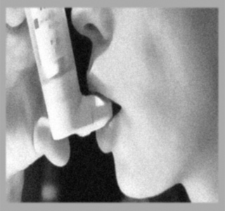
Inhalers for asthma come in many shapes. Most are sprays (Metered Dose Inhalers) and some use powder (Dry Powder Inhalers). A spacer or a holding chamber makes it easier to use a spray inhaler. Asthma medicine also comes as pills and syrups.
The important points which need to be stressed are that, always have asthma medicines, always keep a part of your monthly budget for your medicines and buy medicine well in advance before you run out of medicines. Always carry your quick-relief asthma medicine with you when you leave home.
Risk factors:
Many things can start asthma attacks. These things are called “ risk  factors”: Animals ,Cigarette ,Smoke, Dust in beds with fur, Dust from Strong Pollen from the weather, sweeping smells and trees and  sprays ,flowers ,Colds ,Running, sports, and working hard.
Different people with asthma respond to different risk factors. Know which ones start asthma attacks for you. Keep risk factors that start your asthma attacks out of your home.
The following are some household measures that could help the asthmatic patients.
• Many people with asthma are allergic to animals with fur.  Keep animals outside. Give away pets.
• No smoking inside. Get help to quit smoking.
• Keep strong smells out of the home.  No soap, shampoo, or lotion that smells like perfume. No incense. Make special changes to the room where the person with asthma sleeps.
• Take out rugs and carpets. They get dusty and moldy.
• Take out soft chairs, cushions and extra pillows. They collect dust.
• Do not let animals on the bed or in the bedroom.
• No smoking or strong smells in the bedroom.
•Keep the bed simple. 
Dust collects in the mattress, blankets and pillows. This dust bothers most people with asthma.
• Put special dust-proof covers with zippers on the mattress and pillow.
• Do not use a pillow or a mattress made of straw.
• A simple sleeping mat may be better than a mattress.
• Wash sheets and blankets often in very hot water.  Dry them in the sun.
Use windows to keep the air fresh and clean.
• Open windows wide when it is hot or stuffy, when there is smoke from cooking, and when there are strong smells.
• If you heat with wood or kerosene, keep a window open a little to get rid of fumes.
• Close windows when the air outside is full of exhaust from cars, pollution from factories, dust, or pollen from flowers and trees.
Plan to do these chores when the person with asthma is not there:
• Sweep, vacuum, or dust
• Paint
• Spray for insects
• Use strong cleaners
• Cook strong smelling foods.
• Air out the house before the person with asthma returns.
• If there is no one to help, people with asthma can use a mask or scarf when they sweep or dust.
Running, sports, or working hard can also cause asthma symptoms. But these activities are good for you and need not be stopped. Your doctor may tell you to take asthma medicine before doing these activities. Thus the quality of life need not be affected.
When you know there is asthma in the family, you may be able to keep your baby from getting asthma.
• When you are pregnant, do not smoke.
• Keep tobacco smoke away from the baby and out of your home.
• Put a special dust-proof cover on the baby’s mattress. 
• Keep cats and other animals with fur out of your home.
Health check ups:
Go to the doctor 2 or 3 times a year for check- ups. Go even if you feel fine and have no breathing problems.
• Tell the doctor about any problems with your asthma medicines. The doctor can change the asthma medicine or change how much you take. There are many asthma medicines.
• Ask questions. Your doctor is your partner in controlling your asthma.
• Asthma may get better or it may get worse over the years. Your doctor may need to change your asthma medicines.
Know the signs that your asthma is getting worse and how to respond.
• Be alert for asthma symptoms: Cough, wheeze, tight chest and waking up at night. Act fast if an asthma attack starts.
• Move away from the risk factor that started the attack.
• Take a quick-relief asthma medicine.
• Stay calm for 1 hour to be sure breathing gets better.
Get emergency help from a doctor if you do not get better. Get help if you see any of these asthma danger signs.
• Your quick-relief medicine does not help for very long or it does not help at all. Breathing is still fast and hard.  
• It is hard to talk.
• Lips or fingernails turn grey or blue.
• The nose opens wide when the person breathes.
• Skin is pulled in around the ribs and neck when the person breathes.
• The heartbeat or pulse is very fast.
• It is hard to walk.
Be careful!  Using too much quick- relief medicine for asthma attacks can hurt you.
Quick-relief medicine for asthma makes you feel better for a little while.  It may stop the attack. With some attacks, you may think you are getting better but the airways are getting more and more swollen. Then you are in danger of having a very bad asthma attack that could kill you. A bad asthma attack may be recognized;
• If you use quick-relief medicine more than twice a week to stop asthma attacks, this means you need a preventive medicine for asthma. 
• If you need quick-relief medicine more than 4 times in 1 day to stop asthma attacks, you need help from a doctor today.
Monitoring asthma:
A peak flow meter can be used at a clinic or at home to measure how well a person is breathing.
• It helps the doctor decide if someone has asthma.
• It helps to see how bad an asthma attack is.
• It helps the doctor see how well asthma is controlled over time.
If a peak flow meter is used every day at home, people can find breathing problems even before they start to wheeze or cough.  Then people know when more asthma medicine is needed. There are many kinds of peak flow meters (Consult your doctor for details on this).
Thus on this world asthma day let us all resolve to spread this awareness about asthma and let everyone know that "You Can Control Your Asthma".

Monday, February 15, 2010

The Pickwickian syndrome

Today we look into an interesting syndrome in which obesity decreases ventilation!


The pickwickian syndrome, or obesity hypoventilation syndrome, was first described by Burwell and associates in 1956 and named for a character described in Charles Dickens' The Posthumous Papers of the Pickwick Club. Marked obesity, somnolence, cyanosis, periodic breathing, secondary erythrocytosis, and right ventricular heart failure were the initially described clinical characteristics of this syndrome.
Morbid obesity decreases total lung capacity (TLC), functional residual capacity (FRC), and tidal volume because of increased adipose tissue in the chest wall and elevation of the diaphragm caused by increased intra-abdominal adipose tissue. The reduced lung volumes lead to atelectasis, which alters ventilation-perfusion (V/Q) matching and causes hypoxia. There is also evidence that respiratory muscles become less effective in morbid obesity. Most patients also have OSA, which further increases ventilatory work in a system already predisposed to hypoventilation. Finally, hypoxic and hypercapnic responses are diminished in most patients, either secondary to chronic hypoxia and hypercapnia or, perhaps, congenitally. Even though the exact sequence of events is not always clear, it is easy to conceptualize that affected patients with reduced lung volumes, atelectasis, noncompliant chest walls, ineffective respiratory muscles, upper airway obstruction,and altered metabolic control of breathing are prone to develop hypoventilation, especially during sleep.

Treatment:
Nasal continuous positive airway pressure (nCPAP) is now clearly established as the most effective therapy for OSA. nCPAP is also effective in the majority of patients with obesity hypoventilation, not only resolving upper airway obstruction during sleep, but also increasing the ventilatory response to CO2 while awake and improving awake hypercapnia. However, there are many patients with severe OSA and hypercapnia who are only partially responsive to CPAP alone and continue to demonstrate sleep associated hypoventilation and elevated PaCO2 during wakefulness.

Friday, January 29, 2010

Yousaytoo Awards - a bit offtopic! to break the monotony.

Friends today I am going a bit off topic in my bid to break the monotony and also put forward my optimistic side!
I am extremely glad to share information regarding the "yousaytoo awards". This award is given in the category of internet blogs. I am extremely hopeful and the optimism in myself comes to its its full bloom , the moment i think of a $1,000 holiday dream gift coming my way. It could be a clinic utility with which I could serve my patients.I being a Chest Physician a simple spirometer would be a dream gift for me which I could utilise for my patients. The fun gifts are really funny and the one that interests me the most is the digital photoframe keyring to put the keys of my two wheeler and carry my most memorable pics with me.I know that there are many competitors in the competition and my chances are a lot dependent on luck. Still I am an optimistic person and will continue to enrich my blog with some very useful sites that will definitely be resourceful to everyone.

Sunday, November 22, 2009

Occupational Lung Diseases

 Friends today we will touch one of the very important aspects concerning the respiratory health -- The Occupational Lung Diseases!


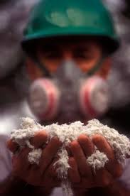
There are various occupational lung diseases. Among them are: Asbestosis, a chronic, progressive inflammation of the lung; it is a consequence of prolonged exposure to large quantities of asbestos, a material once widely used in construction, insulation, and manufacturing. It is not contagious.
Black lung disease is the common name for coal workers' pneumoconiosis (CWP)or anthracosis, a lung disease of older workers in the coal industry, causedby inhalation, over many years, of small amounts of coal dust.
Silicosis is a progressive disease that belongs to a group of lung disorderscalled pneumoconioses. Silicosis is marked by the formation of lumps (nodules) and fibrous scar tissue in the lungs. It is the oldest known occupational lung disease, and is caused by exposure to inhaled particles of silica, mostlyfrom quartz in rocks, sand, and similar substances.
When asbestos is inhaled, fibers penetrate the breathing passages and irritate, fill, inflame, and scar lung tissue. In advanced asbestosis, the lungs shrink, stiffen, and become honeycombed (riddled with tiny holes).
Legislation has reduced use of asbestos in the United States, but workers whohandle automobile brake shoe linings, boiler insulation, ceiling acoustic tiles, electrical equipment, and fire-resistant materials are still exposed tothe substance. Asbestos is used in the production of paints and plastics. Significant amounts can be released into the atmosphere when old buildings or boats are razed or remodeled.
Asbestosis is most common in men over 40 who have worked in asbestos-relatedoccupations. Smokers or heavy drinkers have the greatest risk of developing this disease. Between 1968 and 1992, more than 10,000 Americans over the age of 15 died as a result of asbestosis. Nearly 25% of those who died lived in California or New Jersey, and most of them had worked in the construction or shipbuilding trades.
The risk of having black lung disease is directly related to the amount of dust inhaled over the years; the disease typically affects workers over age 50.Its common name comes from the fact that the inhalation of heavy deposits ofcoal dust makes miners lungs look black instead of a healthy pink. Althoughpeople who live in cities often have some black deposits in their lungs frompolluted air, coal miners have much more extensive deposits.
In the years since the federal government has regulated dust levels in coal mines, the number of cases of black lung disease has fallen sharply. Since theFederal Coal Mine Health and Safety Act of 1969, average dust levels have fallen from 8.0 mg. per cubic meter to the current standard of 2.0 mg. per cubic meter. The 1969 law also set up a black lung disability benefits program tocompensate coal miners who have been disabled by on-the-job dust exposure.
Despite the technology available to control the hazard, however, miners stillrun the risk of developing this lung disease. The risk is much lower today,however; fewer than 10% of coal miners have any x ray evidence of coal dust deposits. When there is such evidence, it often shows up as only small black spots less than 1 cm. in diameter, and may have been caused by smoking ratherthan coal dust. This condition is called "simple CWP" and does not lead to symptoms or disability.
It is estimated that there are 2 million workers in the United States employed in occupations at risk for the development of silicosis. These include miners, foundry workers, stonecutters, potters and ceramics workers, sandblasters, tunnel workers, and rock drillers. Silicosis is mostly found in adults over40. It has four forms:
  • Chronic silicosis may take 15 or more years ofexposure to develop. There is only mild impairment of lung functioning. Chronic silicosis may progress to more advanced forms.
  • Patients with complicated silicosis have noticeable shortness of breath, weight loss, and extensive formation of fibrous tissue (fibrosis) in the lungs. These patients areat risk for developing tuberculosis (TB).
  • Accelerated silicosis appears after 5-10 years of intense exposure. The symptoms are similar to those ofcomplicated silicosis. Patients in this group often develop rheumatoid arthritis and other autoimmune disorders.
  • Acute silicosis develops withinsix months to two years of intense exposure to silica. The patient loses a great deal of weight and is constantly short of breath. These patients are at severe risk of TB.
Occupational exposure is the most common cause of asbestosis, but the condition also strikes people who inhale asbestos fiber or who are exposed to wasteproducts from plants near their homes. Family members can develop the diseaseas a result of inhaling particles of asbestos dust that cling to workers' clothes.
It is rare for asbestosis to develop in anyone who hasn't been exposed to large amounts of asbestos on a regular basis for at least 10 years. Symptoms ofthe disease do not usually appear until 15-20 years after initial exposure toasbestos.
The first symptom of asbestosis is usually shortness of breath following exercise or other physical activity. The early stages of the disease are also characterized by a dry cough and a generalized feeling of illness.
As the disease progresses and lung damage increases, shortness of breath occurs even when the patient is at rest. Recurrent respiratory infections and coughing up blood are common. So is swelling of the feet, ankles, or hands. Other symptoms of advanced asbestosis include chest pain, hoarseness, and restless sleep. Patients who have asbestosis often have clubbed (widened and thickened) fingers. Other potential complications include heart failure, collapsed (deflated) lung, and pleurisy (inflammation of the membrane that protects thelung).
Since the particles of fine coal dust, which a miner breathes when he is in the mines, cannot be destroyed within the lungs or removed from them, builds up. Eventually, this build-up causes thickening and scarring, making the lungsless efficient in supplying oxygen to the blood.
The primary symptom of the disease is shortness of breath, which gradually gets worse as the disease progresses. In severe cases, the patient may developcor pulmonale, an enlargement and strain of the right side of the heart caused by chronic lung disease. This may eventually cause right-sided heart failure.
Some patients develop emphysema (a disease in which the tiny air sacs in thelungs become damaged, leading to shortness of breath, and respiratory and heart failure) as a complication of black lung disease. Others develop a severetype of black lung disease called progressive massive fibrosis, in which damage continues in the upper parts of the lungs even after exposure to the dusthas ended. Scientists aren't sure what causes this serious complication. Somethink that it may be due to the breathing of a mixture of coal and silica dust that is found in certain mines. Silica is far more likely to lead to scarring than coal dust alone.
The precise mechanism that triggers the development of silicosis is still unclear. What is known is that particles of silica dust get trapped in the tinysacs (alveoli) in the lungs where air exchange takes place. White blood cellscalled macrophages in the alveoli ingest the silica and die. The resulting inflammation attracts other macrophages to the region. The nodule forms when the immune system forms fibrous tissue to seal off the reactive area. The disease process may stop at this point, or speed up and destroy large areas of the lung. The fibrosis may continue even after the worker is no longer exposedto silica.
Early symptoms of silicosis include shortness of breath after exercising anda harsh, dry cough. Patients may have more trouble breathing and cough up blood as the disease progresses. Congestive heart failure can give their nails abluish tint. Patients with advanced silicosis may have trouble sleeping andexperience chest pain, hoarseness, and loss of appetite. Silicosis patients are at high risk for TB, and should be checked for the disease during the doctor's examination.
Screening of at-risk workers can reveal lung inflammation and lesions characteristic of asbestosis. Patients' medical histories can identify occupations,hobbies, or other situations likely to involve exposure to asbestos fibers.
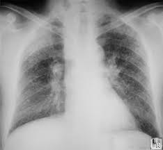
X rays can show shadows or spots on the lungs or an indistinct or shaggy outline of the heart that suggests the presence of asbestosis. Blood tests are used to measure concentrations of oxygen and carbon dioxide. Pulmonary functiontests can be used to assess a patient's ability to inhale and exhale, and acomputed tomography scan (CT) of the lungs can show flat, raised patches associated with advanced asbestosis.
Black lung disease can be diagnosed by checking a patient's history for exposure to coal dust, followed by a chest x-ray to discover if the characteristicspots in the lungs caused by coal dust are present. A pulmonary function test may aid in diagnosis.
X rays can detect black lung disease before it causes any symptoms. If exposure to the dust is stopped at that point, progression of the disease may be prevented.
Diagnosis of silicosis is based on:


  • A detailed occupational history.
  • Chest x rays will usually show small round opaque areas in chronic silicosis. The round areas are larger in complicated and accelerated silicosis.
  • Bronchoscopy.
  • Lung function tests.
It should be noted that the severity of the patient's symptoms does not always correlate with x-ray findings or lung function test results.
The goal of treatment is to help patients breathe more easily, prevent coldsand other respiratory infections, and control complications associated with advanced disease. Ultrasonic, cool-mist humidifiers or controlled coughing canloosen bronchial secretions.
Regular exercise helps maintain and improve lung capacity. Although temporarybed rest may be recommended, patients are encouraged to resume their regularactivities as soon as they can.
Antibiotics may be prescribed to combat infection. Aspirin or acetominophen (Tylenol) can relieve minor discomfort and bronchodilators that are swallowed or inhaled can relax and widen breathing passages.
Diuretics (drugs that increase urine production and excretion) or digitalis glycoside (Digitalis purpurea) are prescribed for some patients. Othersmay need to use supplemental oxygen or use less salt.
Anyone who develops symptoms of asbestosis should see a family physician or lung disease specialist. A doctor should be notified if someone who has been diagnosed with asbestosis:
  • Coughs up blood
  • Continues to lose weight
  • Is short of breath
  • Has chest pain
  • Develops asudden fever of 101°F (38.3°C) or higher
  • Develops unfamiliar, unexplained symptoms.
There is no treatment or cure for black lung disease, although it is possibleto treat complications such as lung infections and cor pulmonale. Further exposure to coal dust must be stopped.
Likewise, there is no cure for silicosis. Therapy is intended to relieve symptoms, treat complications, and prevent respiratory infections. It includes careful monitoring for signs of TB. Respiratory symptoms may be treated with bronchodilators, increased fluid intake, steam inhalation, and physical therapy. Patients with severe breathing difficulties may be given oxygen therapy orplaced on a mechanical ventilator. Acute silicosis may progress to complete respiratory failure. Heart-lung transplants are the only hope for some patients.
Patients with silicosis should call their doctor for any of the following symptoms:
  • Tiredness or mental confusion
  • Continued weight loss
  • Coughing up blood
  • Fever, chest pain, breathlessness, or new unexplained symptoms.
Patients with silicosis should be advised to quit smoking, prevent infections by avoiding crowds and persons with colds or similar infections, andreceive vaccinations against influenza and pneumonia. They should be encouraged to increase their exercise capacity by keeping up regular activity, and tolearn to pace themselves with their daily routine.
Asbestosis can't be cured, but its symptoms can be controlled. Doctors don'tknow why the health of some patients deteriorates and the condition of othersremain the same, but believe the difference may be due to varying exposuresof asbestos. People with asbestosis who smoke, particularly those who smoke more than one pack of cigarettes each day, are at increased risk for developing lung cancer and should be strongly advised to quit smoking.
Those miners with simple CWP can lead a normal life. However, patients who develop black lung disease at an early age, or who have progressive massive fibrosis, have a higher risk of premature death.
Silicosis is currently incurable. The prognosis for patients with chronic silicosis is generally good. Acute silicosis, however, may progress rapidly to respiratory failure and death.
Workers in asbestosis-related industries should have regular x rays to determine whether their lungs are healthy. A person whose lung x ray shows a shadowshould eliminate asbestos exposure even if no symptoms of the condition haveappeared.


Anyone who works with asbestos should wear a protective mask or a hood with aclean-air supply and obey recommended procedures to control asbestos dust. Anyone who is at risk of developing asbestosis should:
  • Not smoke
  • Be vaccinated against influenza and pneumonia
  • Exercise regularlyto maintain cardiopulmonary fitness
  • Avoid crowds and people who haverespiratory infections.
A person who has asbestosis should exercise regularly, relax, and conserve energy whenever necessary.
The only way to prevent black lung disease is to avoid long-term exposure tocoal dust. Coal mines may help prevent the condition by lowering coal dust levels and providing protective clothes to coal miners.
Silicosis is a preventable disease. Preventive occupational safety measures include:
  • Controls to minimize workplace exposure to silica dust
  • Substitution of substances--especially in sandblasting--that are less hazardous than silica
  • Clear identification of dangerous areas in the workplace
  • Informing workers about the dangers of overexposure to silica dust, training them in safety techniques, and giving them appropriate protectiveclothing and equipment.
Coworkers of anyone diagnosed with silicosis should be examined for symptomsof the disease. The state health department and the Occupational Safety and Health Administration (OSHA) or the Mine Safety and Health Administration (MSHA) must be notified whenever a diagnosis of silicosis is confirmed.
http://www.faqs.org/

Promote your blog

Promote Your Blog